
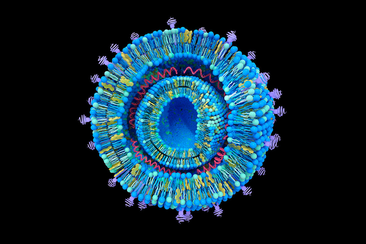
Nucleic Acid LNP Using Microfluidics

For mRNA, and some other nucleic acid therapeutics, formulation development involves encapsulating the nucleic acid in lipid nanoparticles (LNPs) to provide stability and enhance cell delivery. Formulation is generally performed using a microfluidic process, which enables controlled, reproducible and scalable production of LNPs. Apart from provision of the mRNA, the initial task in LNP formulation development is the selection of the lipid components, particularly the selection of the ionizable/cationic lipid.
Cationic lipoplexe structure. Cationic lipoplexes are one type of Lipid Nanoparticles (LNPs) used for gene delivery. Nucleic acids intercalated between the lipid bilayers. 3d illustration

Key components of LNPs

    Ionizable lipids

    Charge-neutral at physiological pH but become positively charged at acidic pH, enabling mRNA binding and endosomal escape

    Phospholipids

    Stabilize the lipid bilayer

    Cholesterol

    Enhances membrane stability and fluidity

    PEGylated lipids

    Provide a hydrophilic “stealth” coating to improve circulation time and prevent immune recognition

    Additional actives

    Agents to modulate activity or cell-specific delivery

    Optimal LNP formulation development also requires the optimization of the microfluidic process

    • Preparation of mRNA and lipid solutions: selection of the pH and ionic strength of the aqueous buffer which will impact the charge and stability of the mRNA and lipids, providing optimal electrostatic interaction for encapsulation

    • Mixing parameters involving mixer design, flow rate ratio (FRR: ratio of aqueous to ethanolic solution) and total flow rate (TFR)

    • Buffer exchange by dialysis/ultrafiltration to remove solvent and exchange the LNPs into a physiological buffer and sterile filtration

    Illustration of a lipid nanoparticle (LNP) encapsulating mRNA. The central structure is a spherical particle with layered lipid bilayers composed of various molecules. Inside the particle, curly red strands represent “MARV GP sa mRNA” labeled with a 5’ cap (m7Gppp), untranslated regions (UTRs), and a poly-A tail. Five labeled components of the LNP structure are indicated with red arrows: 1. Cholesterol – Shown with a molecular structure and labeled on the left side of the nanoparticle. 2. D-Lin-MC3-DMA (Ionisable lipid) – Positioned on the lower left with its chemical structure. 3. DSPC (1,2-distearoyl-sn-glycero-3-phosphocholine; 18:0/18:0 PC) – Displayed on the upper right. 4. Dimyristoylglycerol-PEG – Shown on the lower right with its molecular structure and PEG tail. 5. MARV GP sa mRNA – Centered inside the nanoparticle with a schematic representation of the mRNA strand. The diagram visualizes how different lipids and cholesterol form a nanoparticle that encapsulates mRNA for delivery, commonly used in mRNA vaccine technology.

    In line with ICH Q8 (R2)(Pharmaceutical Development), ICH Q9(Quality Risk Management), i.e., QbD, Labcorp biopharmaceutical CMC services’ LNP formulation development process incorporates a design of experiment (DoE) statistical approach to evaluate both LNP components and the microfluidic process. Using DoE provides the maximum amount of data for the minimum number of samples and hence also reduces the analytical load. The outcome of these studies is a lead formulation that will meet prescribed critical quality attributes, such as particle size and size distribution, zeta potential, % mRNA encapsulation and mRNA integrity. Real-time, accelerated and forced degradation studies can be applied to determine the formulation provides sufficient stability and expected shelf life.

    Pre-formulation resources

    Article

    Forced degradation of biopharmaceuticals: Protein stress testing

    Info Sheet

    Formulation development: The key to product stability