Posters
Multiplex IHC for patterns of protein expression, co-expression, and spatial relationships
01 Apr 2018
Multiplex immunohistochemistry (IHC) is an assay that utilizes the basic concept of single antigen staining to detect multiple markers at one time. It allows for the labeling capabilities of peroxidase, alkaline phosphatase and other conjugated primary or secondary antibodies and can be achieved using several common methods including indirect or direct staining protocols
Researchers have been directing their focus on multiplex IHC due to the many advantages of using multiple antibodies at once. It allows us to gather higher quantities of data at one time while saving precious tissue samples. Spatial data can be obtained for one protein target relative to another as well as in proportion to tissue architecture or organelles that are nearby. It is often used to determine subsets of immuno-oncology markers in tumors, telling us a more in-depth story of what is happening in the tumor environment. Multiplex IHC has also become an important solution for determining the co-expression of markers in cells.
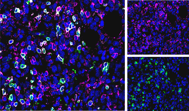
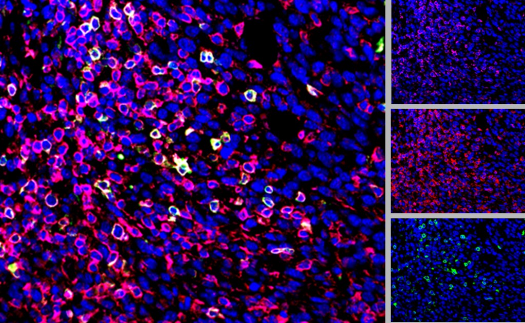
Fig 2: CD3 (pink), CD4 (green), CD45 (red), and DAPI (blue) Staining in CT26 tumor.
Labcorp is now offering up to four-color multiplexing with conjugated antibodies and is continuously striving to validate new multiplexing options such as chromogen detection. Optimized protocols have been developed on the Bond RXm Autostainer for consistent staining and on the Aperio VERSA Scanner to obtain high-resolution images of the data.
Contact us to learn more about custom marker requests for multiplex IHC as well as our other IHC services.
Note: Please note that all animal care and use was conducted according to animal welfare regulations in an AAALAC-accredited facility with IACUC protocol review and approval.