
Poster
Luciferase-labeled cell lines: a valuable tool for oncology studies
In preclinical research, small animal in vivo imaging plays an essential role in visualizing physiological processes, progression of disease and development of therapies. Luciferase (luc) enabled cell lines offer a simple, high-throughput and robust means to quantitatively assess tumor burden and response of tumors to treatment therapies in subject animals, through bioluminescence imaging (BLI)1, 2. Click here to learn more about BLI
The sensitivity and accuracy of BLI systems offers multiple advantages over traditional methods, such as:
(1) Noninvasive real-time whole-body in vivo tumor monitoring and imaging
(2) Continuous assessment of tumor progression and response to therapeutic treatments in the same animal
(3) Metastasis assessment
(4) Reduced need for animal sacrifice
Why Labcorp Drug Development (formerly Covance Laboratories)?
Covance was the first Contract Research Organization (CRO) to offer BLI, in 2003, and in the past 18 years we have accrued extensive experience in this optical imaging field. We offer a large panel of luciferase-enabled cell lines with over 80 unique hematological malignancy cell lines (Table 1). We have a dedicated team of experts to design the best oncology study for you as well as to ensure smooth study execution. Our scientists are skilled to engineer our in-house cell lines or your cell line of interest as well as to custom make vectors to express luciferase for BLI detection. Complete list of cell lines
LabCorp Drug Development (formerly Covance Laboratories) has a license agreement from Dana Farber Cancer Institute and other organizations that provides additional access to many characterized, in vivo validated luciferase-expressing tumor lines.
Our luciferase-expressing tumor cell lines have
· Stable luciferase expression
· A fluorescent protein (mCherry) along with the Luc 2 gene
· Quantitative correlation between signal strength and cell numbers
· High sensitivity and low signal-to-noise ratio
· Availability of multiple tumor cell lines from human, mouse, and rat
· Suitable for in vitro as well as for in vivo assays
We also offer an alternative method for cell transduction in luc- cell enabling which is cell electroporation allowing clients to choose customized vectors instead of a lentiviral vector.
Service offerings related to BLI include:
- Cancer Tumor Models
- Radiation Therapies
- Flow Cytometry
- Immuno-Oncology & Immunotherapy
- CAR T cell persistence
- ABSL2 studies
How it works?
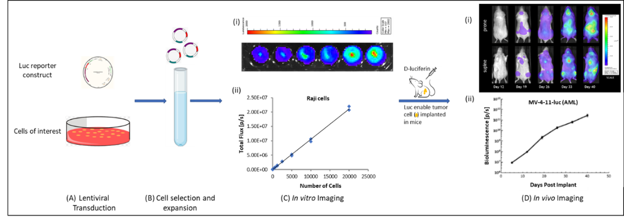
Typically, cancer cells are engineered to express the firefly luciferase gene along with a puromycin resistance gene using a lentiviral system for transduction (Fig. 1A). Along with the Luc 2 gene, the construct has a fluorescent protein coding gene (mCherry) that enables the detection of tumor cells in different tissues over time. Cells are cultured in the presence of puromycin to select the cells with inserted lentiviral vector encoding firefly luciferase (Fig. 1B). Light output is generated (Fig. 1C(ii)) from the luciferase enabled cells and bioluminescence is measured using the IVIS® In Vivo Imaging System (Fig. 1C (i)); Luc-enabled cells are then engrafted into mice to form tumors. Following the injection of D-luciferin (substrate), the luciferase enzyme will catalyze this substrate resulting in light emission detected with IVIS® (PerkinElmer, Waltham, MA) (Fig. 1D(i)) and analyzed in regions of interest using the Perkin Elmer's Living Image software (Fig. 1D (ii)).
Contact us to request the full data set or to learn more about our luciferase enabling service and how it can be applied to your preclinical research.