
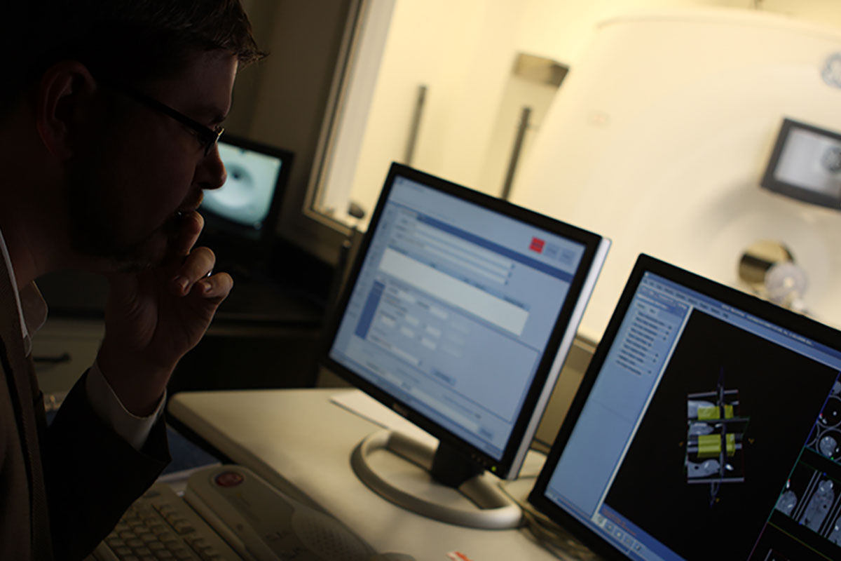
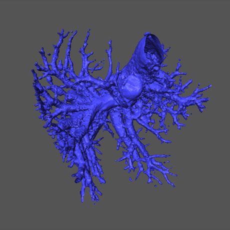
Computed Tomography (CT)

Micro computed tomography (CT) imaging platform draws on expertise in implementing, developing, optimizing, and validating micro-CT imaging paradigms for drug research studies. Our experience in CT-based bone and soft tissue imaging encompasses a number of disease states.

CT-based bone and soft tissue imaging

In vivo micro-CT imaging enables high-resolution (to ~50 microns), three-dimensional image generation by rotation of an x-ray source and detector around an imaging subject. Multiple projections are obtained during the rotation and reconstructed to generate the three-dimensional image. The large density differences between bone, soft tissue, and air make them readily distinguished by CT. CT contrast between different soft tissues is generally more subtle. The use of systemically administered, CT contrast agents enables better visualization of the soft tissues, including the vascular system.

Mouse liver ex vivo after perfusion with a high density fixing/curing agent

Bone Imaging and Metastasis

CT is the preferred, translational technology for the quantification of metastatic and arthritic bone disease. We provide numerous, validated bone-disease models, including several models of bone metastasis and rheumatoid arthritis. Micro-CT can be used to characterize:

  • Bone loss and growth

  • Bone density and microstructure

  • Bone remodeling and calcification

We also have strong expertise in imaging of bone metastasis in validated intracardiac and peritibial models, including imaging of osteolytic and osteoblastic activity, using staged-treatment or prevention paradigms.

Tumor Volume Quantification